
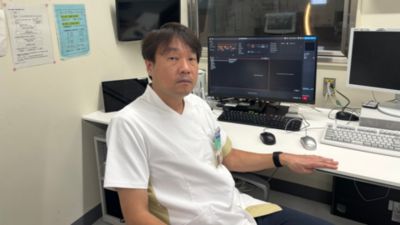
Hideto Miyano is Chief Technologist (Engineer) of Toyonaka Municipal Hospital Central Medical Bureau Radiology Department.
Opting for the SmartPath to Evolution upgrade for their Ingenia 1.5T instead of purchasing a new system, has brought the latest technology and appearance to the MRI system at Toyonaka Municipal Hospital. Choosing this upgrade helped the hospital in cutting costs, reducing downtime and enhancing their patient‐centered environment. The MRI team now experiences remarkable improvements in workflow, benefiting patients and staff. Excellent diagnostic images are acquired, while efficiency in handling patients and performing scanning are achieved.
Hideto Miyano is Chief Technologist (Engineer) of Toyonaka Municipal Hospital Central Medical Bureau Radiology Department.
Toyonaka Municipal Hospital in Japan performs approximately 6,500 MRI examinations per year with a main focus on head, vertebral bodies, limb joints, upper abdomen and lower abdomen, mainly on pediatric patients. It is a designated secondary emergency hospital, and its MRI room accepts stroke patients 24 hours a day.
The hospital used to operate two 1.5T MRI machines until it was decided to replace the Achieva 1.5T with a Philips Elition 3.0T system. The recent upgrade of the hospital’s Ingenia 1.5T with SmartPath to Evolution has brought latest technology and appearance to that system and the MRI team experiences remarkable improvements in workflow and clinical usefulness.

Toyonaka Municipal Hospital (Toyonaka City, Osaka Prefecture) is a general hospital and designated as a regional base hospital for cancer treatment cooperation. It provides safe, high‐quality medical care and strives to be a warm‐hearted hospital that takes the patient’s perspective into account.
When the Ingenia 1.5T had been in operation for approximately nine years, the hospital was considering options for updating or replacement. “At the perfect time, we received a proposal for SmartPath to Evolution 1.5T,” says Hideto Myano. This upgrade brings both technology and appearance to the latest systems’ level, while making use of the existing magnet.
“Benefits of this approach include reduced installation costs and limited downtime. Being able continue use of our existing magnet, but with an updated technology level, was an important reason why we chose SmartPath to Evolution 1.5T. And operating two MR systems having the same modern user interface, will make it easier for operators to work alternately on both systems.”

SmartPath to Evolution 1.5T.
"Emphasis often seems to be on obtaining high‐quality images in a short time using Philips top quality, high‐speed technology, observes Hideto Miyano.“ However, SmartPath to Evolution 1.5T also comes with the concept of ‘improving the examination environment by putting the patient at the center’. I see it has many technologies that help improve the workflow, which is what clinical sites have been looking for.”
During the scan, patients automatically receive spoken and visual guidance, which often helps alleviate patients’ anxiety. It provides breath holding instructions, allows the patient to see the examination’s progress and remaining scan time and get prompts when the table will move. All of this contributes to patients feeling at ease during the examination.
The comfortable, extra‐thick mattress is designed to reduce the effects of pain and distress even during long examinations. It also helps reduce patient motion that causes motion artifacts, which in turn reduces the risk of needing to redo scans.
SmartPath to Evolution has many technologies that help improve the workflow, which is what clinical sites have been looking for.
Hideto Miyano appreciates how workflow for technologists can be considerably improved, for instance with the interactive touchscreen’s features like automatic patient centering (SmartTouch) during patient setup and automatic start of scanning when the examination room door (SmartStart) is closed. The touchless breathing synchronization device VitalEye is always on, so setting up a breathing sensor is no longer needed, which improves workflow and saves time. Some of these relatively simple innovations can help shorten examination time slots, which is exactly what users have been looking for for years.
“SmartPath to Evolution came with AutoVoice, which was not available on our Ingenia, that not only gives breathholding instructions, but also announces table movements and informs the patient of the remaining scan time, which used to be done manually by the MR technologists,” says Hideto Miyano. “So, these features not only help many patients feel more comfortable during the examination, but also relieves the technologists.”
“SmartPath to Evolution 1.5T has helped us realize the importance of improving patient‐centered workflows and has allowed us to focus on acquiring high‐quality diagnostic images, which is a priority in our hospital,” says Hideto Miyano.
Patients can undergo examinations with peace of mind and comfort, and the introduction of the Smart Workflow solution has shortened the time it takes to set up patients, allowing the Toyonaka technologists to focus on patient care more than ever before and perform examinations with more time to spare.
SmartPath to Evolution 1.5T helped us realize the importance of improving patient‐centered workflows and has allowed us to focus on acquiring high-quality diagnostic images, which is a priority in our hospital.
“These improvements have allowed us to add sequences tailored to initial clinical findings, without exceeding the examination time,” says Hideto Miyano. “Adding sequences like NerveVIEW, FRACTURE and DWI TSE XD enhance our clinical confidence, helping obtain diagnostic examinations without compromise. In terms of image quality, beautiful images with high SNR, high resolution and stable fat suppression have been obtained. SmartPath to Evolution 1.5T has improved our patient-centered examination environment and provides high‐quality, clinically useful images.”

Routine Cervical spine imaging (DWI, 3D NerveVIEW) in a case of postoperative edema for cervical myelopathy.
The touchless breathing synchronization device VitalEye uses an infrared camera and removes the need for setting up a breathing sensor, which improves workflow and saves time.
Previously, when a respiratory sensor or navigator echo was used, sudden movements such as coughing or irregular breathing cycles affected respiratory gated imaging. Use of VitalEye removes such sudden breathing and its AI‐based respiratory waveform recognition provides highly accurate respiratory‐gated imaging, surpassing conventional methods. “We see improvements in the accuracy of our respiratory gated imaging and a reduced risk of re‐imaging,” says Hideto Miyano. VitalEye can also be applied when imaging the lower abdomen and shoulder joints, expanding the range of imaging methods in those areas.

3D MRCP with VitalEye respiratory gated imaging shows better image quality than respiratory synchronization with traditional breathing sensor, which results in poor image quality due to irregular breathing cycles (left). With VitalEye the effects of irregular movements are reduced (right).
Dr. Hideto Miyano’s first observation about SmartSpeed AI was that high‐quality images can be obtained in short scan times. This is particularly advantageous in single shot sequences with low SNR, and he is actively applying it, mainly in abdominal imaging. “We can now obtain images with high SNR at around four times the speed, while reducing the effects of blurring.”

Routine fetal imaging using single shot TSE, SmartSpeed AI and SmartSpeed DWI. Single shot TSE does not use halfscan and produces sharp images with high SNR.
"Many head and abdominal examinations are performed at our hospital, and our examination times have now been reduced by about four to five minutes. Specifically for routine head imaging in emergency patients, the total imaging time has been considerably shortened to less than a minute, and our way of working for emergency examinations has been improved."
Specifically for routine head imaging in emergency patients, the total imaging time has been considerably shortened to less than a minute, and our way of working for emergency examinations has been improved.

Acute head imaging using SmartSpeed. Total imaging time was 3:56 min.
Since the introduction of SmartSpeed AI, we can now perform examinations faster and with more flexibility.
Previously, the MR team was often swamped with examinations, Dr. Hideto Miyano explains. “However, since the introduction of SmartSpeed AI, we can now perform examinations faster and with more flexibility. For instance, when our 3.0T system was unavailable for half a day, we managed to complete all examinations by using the 1.5T system. Thanks to SmartSpeed's capability to shorten scan times while providing high image quality."

MRI technologists Nakai (left) and Miyano (right).
In areas that are often affected by body movements, like the shoulder joints and the pelvic cavity, Dr. Hideto Miyano uses SmartSpeed MotionFree. It leads to reproducible examinations and helps him reduce the need for re‐imaging because of motion artifacts.
He values that SmartSpeed MotionFree images provide a natural appearance in body imaging, without contrast loss and chemical shift resembling Cartesian imaging. Furthermore, the FOV change from circular to rectangular benefits the body motion correction technology for him.

Routine MRI of shoulder with SmartSpeed MotionFree.

mDIXON XD FFE and 3D VANE XD MRA. By combining the DIXON method and body motion correction technology, the three branches from Aorta are clearly visualized. Acquired voxels are 1.39 x 1.39 x 1.8 mm, scan time is 1:13 min per station.
Dr. Hideto Miyano observed improved image uniformity in large field-of-vies (FOV) imaging, thanks to improvements in static magnetic field uniformity and uniformity correction since SmartPath to Evolution 1.5T. He notes that joint unevenness and blurring in multi‐station imaging, such as total spine and DWIBS, are notably reduced compared to their imaging before SmartPath to Evolution.

Total spine and DWIBS imaging showing multiple bone metastases in a lung cancer patient.
In the past, additional stations were sometimes scanned when inhomogeneity and blurring arose near the overlap, particularly in total spine imaging. However, with SmartPath to Evolution, excellent quality is obtained in all stations and the overlap has been reduced, allowing for a wider imaging coverage than previously in their Ingenia 1.5T. This has improved their workflow and efficiency.

Comparison of total spine multi‐station imaging. No effects of unevenness or blurring at seams are observed with SmartPath to Evolution 1.5T. Moreover, the overlap of stations was reduced from 80 mm to 40 mm.
